In this 5 part series we will discuss a very important and at times overlooked area of residential water treatment, the Disinfection and Sanitization for Residential Water Treatment. The information in this series was prepared by Larry Zinser of Master Water Conditioning. Larry Zinser is a sales engineer with Master Water Conditioning. Following an education with a Bachelor's Degree in Chemistry from Georgetown University, Larry spent 27 years in the Marine Corps before retiring as a Colonel. Since then, Larry has built his career designing, building, teaching and troubleshooting water treatment systems and high purity water applications. His background is in commercial, industrial, and residential applications and he has provided accredited technical courses throughout the country and internationally.
This discussion of disinfection for residential applications will include five steps. First, we will look at the species that pose a threat to human health. Second, we will look at where the threat comes from, and how it can occur and then reoccur. Third, we will look at the overall strategy, which we as service technicians can take to combat and minimize the threat. Fourth, we will then look at the tactics and specific actions which should be taken to combat microorganisms. Finally, we will look at four common installation types, and the practices we can take to win the battle against microbiology.
Part 1: The Threat
These four microbiological species are the most serious threats in water.
-
Bacteria
Bacteria represent the first forms of life on our planet. They exist in every habitat known on earth from the extreme cold of the arctic to the extreme heat of sulfur springs. There are about 2.5 million bacteria in every gram of fertile soil and 1 million in every milliliter of natural water. Their size ranges from a third of a micron to as large as 750 microns. A human hair is about 10-40 microns in diameter. Most bacteria are in a size range of ½-micron to 5 microns.
Bacteria reproduce by binary fission (they split in two), and tend to form into colonies when provided with nutrients and a solid surface. Most disease-causing bacteria live off of other organisms. E. coli are rod-shaped bacteria that use a flagella or tail to move. They live in the lower intestines of warm-blooded animal, but can live for short periods outside of the human body. Some species of E. coli can cause food poisoning, but most species are harmless. The lower intestines are also inhabited by other more toxic organisms (called pathogens). E. Coli and some of its close relatives are referred to as fecal coliform. They are relatively easy to detect in on-site testing so their presence in testing is used primarily as an indicator of the other more toxic inhabitants of the lower intestines.
Mycobacterium Tuberculosis is a spherical and pathogenic bacteria. It is aerobic, meaning that it requires an abundance of oxygen, which it finds in the human lungs. It is spread from lungs to lungs. Salmonella is a rod shaped bacteria with flagella for motion. It is carried by animals, milk products, reptiles and tainted fruits and vegetables. It causes typhoid fever and Salmonellosis. Pseudomonas aeruginosa is found in soil and in water, and can cause severe inflammation of all parts of the human body, especially the lungs, kidneys and urinary tract. The cell surface contains polysaccharides which allow it to form biofilm. Shigella is also a rod-shaped bacteria that causes dysentery and shigellosis. In Figure 1, those outlined in blue are typically found in water.
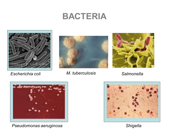
Figure 1. Images of common bacteria associated with disease and illness.
-
Virus
Viruses are extremely small infectious agents which are found in every ecosystem on earth. It is the most abundant type of biological entity. Their shape ranges from helical to icosahedral (20-sided hedron) Viruses can only replicate within living cells. Many are carried by vectors, bacteria, and through the air in coughs and sneezes. The rotovirus and norovirus are essentially a double strand of RNA within a shperical shape. They is found in the small intestines where it replicates in the cell walls of the intestine lining. They are transmitted by fecal-oral route and enter the water stream embedded in bacteria. Rotovirus is the leading cause of severe diarrhea and “stomach flu” in small children. With age, the human body develops an immunity to this virus. Norovirus is responsible for 90% of the non-bacterial outbreaks of gastroenteritis (“gastric flu”) around the world. Outbreaks often occur in semi-closed communities, such as long term care facilities, hospitals and cruise ships. Varicella Zoster virus causes the highly contagious Chicken Pox. It is spread through coughing or sneezing. Normally, a life-long immunity is developed after infection, but it can sometimes reoccur decades later as shingles. The Adenovirus is a icosahedral or 20-faced hedron shaped virus consisting in a double strand of DNA and other proteins. It is the cause of about 10% of upper respiratory infections in children and in some adults. Influenza is another RNA virus that exists in birds and mammals. It causes symptoms from general discomfort to pneumonia. This is not the “24-hour flu”. It replicates in the nose, throat and lungs of humans and in the intestines of birds and is spread in seasonal epidemics. There are three forms: A is the most virulent form for humans (H1N1 of 2009), and is shared with wild and aquatic birds; B is a human-only virus which is not widely spread; and C is shared by humans, dogs and pigs, but is rare and generally mild in effect.
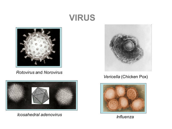
Figure 2. Images of various common viruses that affect human health. Viruses are extremely small in size and can only replicate in other living cells.
-
Protozoa
Protozoa are relatively large, up to 50 microns, they consume biomass and they prey on bacteria. Cryptosporidium is a parasite closely related to algae. They are single-celled protists: mobile, having a cell covering (sometimes a shell) and have a distinct life cycle. They alternate between a proliferate stage when they reproduce, and a cyst (protected) stage when they travel between hosts. They cause acute short-termed infection of human intestines. Cryptosporidium is spread through contaminated water; it generally follows the fecal-oral route. It travels encase in a hardy microbial cyst. Entamoeba are anaerobic parasites that live in the lower intestine. In their life cycle, they form a cyst covering and exit the body with feces. When they re-enter a body and move to the intestines, the cyst dissolves, and they multiply by binary fission. Then each forms its own cyst. They cause dysentery and liver abscess.
Plasmodium is the parasite that causes Malaria. Its life cycle includes a mosquito vector and a vertebrate host. The protozoa is delivered to the host by the bite of the female mosquito. It travels the bloodstream to the liver where it multiplies and may undergo a period of inactivity. Once it leaves the liver, it travels the bloodstream consuming hemoglobin and causing the malaria symptoms. Giardia Lamblia is a flagellated anaerobic protozoa that replicates by binary fission in the small intestines. It lives in humans, dogs, cats, birds beaver and sheep. It is prevalent in ponds with beaver dams. After formed, the protozoa forms a cyst and exits the body in feces. It can live (as a cyst) for weeks to months in cold water. Upon ingestion, it moves to the small intestine to repeat the cycle.
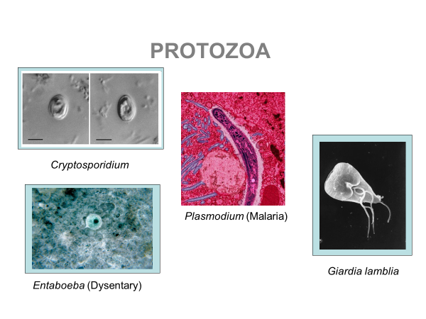
Figure 3. Images of various protozoa. Protozoa are free swimming organisms found in aquatic environments.
-
Fungi
Fungi are mostly multicellular organisms, though some yeast are single celled. Fungi have a complex cellular structure and distinct nuclei with genetic material. Most are tubular or elongated in shape. They are primarily terrestrial, but some are aquatic and others are airborne. There are about 100,000 species identified. The Candida albicans, a form of yeast, can cause oral and genital infections, called yeast infections. It lives in the human mouth and intestines of about 80% of humans as a single cell organism with no harm. Upon an environmental cue, it can turn into a multicellular filament and infect the body. Candida glabrata is also prevalent and generally non-pathogenic. It can form a biofilm on dentures and urinary catheters.
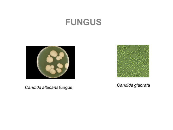
Figure 4. Fungi can range in size from the cellular level to macro organisms like mushrooms
Commensal, Mutual or Parasitic
Not all microorganisms, however, are harmful, or pathogenic. Most, in fact, are harmless, or “Commensal”. E. Coli, for example, as we have already discussed, is normally harmless, and it is useful to us as an indicator of other more dangerous microbes. Humans depend on microorganisms for life, but not directly. Microbes are responsible for nitrogen fixation which converts nitrogen gas to a usable building blocks of life such as proteins and amino acids. However, there are no true “Mutual” relations between the human body and microorganisms. As another example of our use of microbes, there are somewhere between 300 and 1000 different species of microorganisms in the “Gut Flora” most of which help the human body functions. Gut Flora ferment carbohydrates to produce useful calories, and they stimulate lymphoid tissue to produce antibodies to ward off pathogens. There are also “Mutual” relations outside the human body, however, like the use of yeast for fermentation, or the use of mycorrhizal fungus for mushrooms as food for humans.
With infection, numbers do matter. The challenge to health is caused not by single microorganisms, but by colonies of pathogenic organisms. When a pathogen can multiply enough to overwhelm the body’s natural immune system, then there are health issues.